Skip to Content


Crysalin, established in June 2007, focuses on developing a protein lattice technology initially conceptualized in 2002 at the Oxford University Laboratory of Molecular Biophysics.


  • Crysalins are advanced protein-based nanostructures designed for efficient protein structure determination. This technology offers distinct advantages over traditional methods, as it imposes order on macromolecules, enabling X-ray and electron microscopy to resolve 3D molecular structures. This capability is particularly useful for challenging targets, such as membrane proteins and flexible soluble proteins, where conventional techniques often fail. Additionally, crysalins show potential applications in biosensor development and optoelectronics.


  • The company has secured funding from various investors, including IP Group plc, Longwall Venture Partners LLP (Oxford Technology ECF fund), SandAire, Oxford Spinout Equity Management, Ora (Guernsey) Ltd, and the Oxford University Challenge Seed Fund (UCSF).


Exemple : "Visualization of Protein Lattice Structures for Enhanced 3D Molecular Determination"


 Technology


Our technology is based on a new type of highly ordered materials called crysalins, which are created by modifying natural protein building blocks. These crysalin lattices are stable, easily assembled, and have high porosity, allowing for the dynamic incorporation of guest molecules.


Formation of 2D & 3D Crysalins

To aid in the structural analysis of biological macromolecules, Crysalin has developed a novel crystalline material by genetically fusing oligomeric proteins with specific symmetries (as described in the original crysalin patent, GB2393959).


Crysalin lattices are formed from oligomeric proteins with high symmetry, belonging to Tetrahedral (T), Octahedral (O), Dihedral (Dn), and Cyclic (Cn) point groups. These proteins utilize their inherent symmetry to generate lattices through genetic fusion with other proteins that have matching symmetry and coordination properties. Each protein fusion is referred to as a crysalin “protomer.”


We can consistently produce 1D, 2D, and 3D Crysalins.


These engineered lattices are designed to serve as frameworks for the controlled attachment of guest macromolecules. This enables rapid and automated structure determination of macromolecules and their complexes with other macromolecules, small molecule co-factors, substrates, and d rugs using X-ray diffraction or electron microscopy.​

Methods for analyte attachment

Attachment of analyte macromolecules to the crysalin lattice and protocols to array these attached molecules in an identical conformation is essential for the use of crysalins in structure determination.


Covalent attachment of analytes through genetic fusion has been successfully achieved. Alternative approaches include attachment of analyte molecules to a pre-formed lattice via non-covalent interactions.

Elucidation of protein structure

To date we have had success in attaching analytes to 2D and 3D crysalins, allowing observation of bound proteins and determination of protein structure:

 We specialize in deciphering the 3D architecture of proteins and other macromolecules using state-of-the-art techniques such as : 

X-ray Crystallography

X-ray crystallography is a technique used to determine the atomic and molecular structure of a crystal. When X-rays are directed at a crystalline sample, they diffract in specific patterns. By analyzing these diffraction patterns, the electron density distribution within the crystal can be mapped, allowing for the precise determination of the three-dimensional arrangement of atoms within the molecule.

Cryo-Electron Microscopy (Cryo-EM)

Cryo-electron microscopy is a method used to observe biological macromolecules at near-atomic resolution. In this technique, samples are rapidly frozen to preserve their native structure without the need for staining. Electron microscopy is then used to capture high-resolution images, and computational methods are applied to reconstruct 3D models of the macromolecule, providing structural information on molecules that are difficult to crystallize.


Nuclear Magnetic Resonance (NMR) Spectroscopy

NMR spectroscopy is a technique used to investigate the structure and dynamics of molecules, particularly in solution. It relies on the magnetic properties of certain atomic nuclei. When exposed to a magnetic field and radiofrequency radiation, nuclei resonate at specific frequencies. The resulting spectra provide detailed information about the chemical environment, connectivity, and three-dimensional structure of the molecule in solution.


Protein Production


Learn More

Efficient and scalable protein production is at the heart of our technological solutions. We harness cutting-edge bioprocessing techniques, including :

  • Recombinant DNA Technology for producing proteins in various host systems (bacteria, yeast, mammalian cells).
  • Optimizing yield and purity through advanced purification methods such as chromatography and affinity tagging.
  • Scaling up production for therapeutic proteins, enzymes, and vaccines.

    Crystallization of Insulin: Topic Walkthrough

Learn how the process of insulin crystallization works and how it plays a crucial role in both purifying insulin and determining its molecular structure.

Getting Started with Insulin Crystallization

Crystallizing insulin is a straightforward yet highly technical process used in pharmaceutical applications and structural studies. The first step involves preparing an insulin solution by dissolving the hormone in an appropriate solvent, usually water or saline. This solution is then carefully adjusted for temperature, pH, and concentration to ensure optimal crystallization conditions.


Once the solution is ready, crystallization is induced by controlling factors like concentration, temperature, and the addition of precipitating agents, such as salts. These changes cause insulin molecules to self-assemble into regular and ordered crystal structures. This process may take time, but careful monitoring ensures the creation of high-quality crystals.


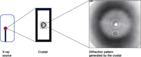
After the crystals form, they are characterized using techniques like X-ray diffraction to determine the three-dimensional structure of insulin. X-ray diffraction provides detailed information about the arrangement of atoms and the formation of disulfide bonds in the insulin molecules.

Read More

Updates and Improvements in Insulin Crystallization

Crystallization techniques are continually evolving, with new methods and improvements in technology helping to refine the process. Researchers are working on developing new strategies for optimizing crystal formation, improving resolution, and reducing the time required for successful crystallization.


Advancements in cryo-crystallography, improved stabilizing agents, and enhanced X-ray diffraction techniques have all contributed to more accurate and efficient insulin crystallization. These improvements ensure better-quality insulin for pharmaceutical production and enable more precise structural analysis of the protein.

Support and Resources for Crystallization Studies

Researchers involved in insulin crystallization have access to numerous resources to aid in their studies. Specialized software and techniques for X-ray crystallography, as well as consultation with experienced crystallographers, help optimize the crystallization process.

Moreover, there are extensive research papers and technical documentation available on crystallization protocols, allowing scientists to refine their methods based on the latest findings and techniques. Regular updates from research institutes and publications also contribute to ongoing improvements in insulin crystallization methods.

These resources and continual research efforts ensure that insulin crystallization remains a reliable and effective process for both drug production and structural analysis.


Protein crystallization for X-ray crystallography